Magnetic Resonance Imaging, or MRI, is a non-invasive medical imaging exam that produces detailed images of almost every internal structure in the human body, including the organs, bones, muscles, and blood vessels. MRI scanners create images of the body using a magnetic field and computer-generated radio waves. MRI can be used to diagnose aneurysms, spinal cord conditions, stroke, tumors, a variety of traumatic injuries and many other diagnosis. Internal organs are often examined through MRI are the liver, kidneys, spleen, pancreas, uterus, ovaries and prostate to name a few. Sometimes contrast agents are used to improve the visibility of internal body structures under magnetic residence imaging. The most commonly used compounds for contrast enhancement are gadolinium-based contrast agents. Most MRIs do not require contrast injection.
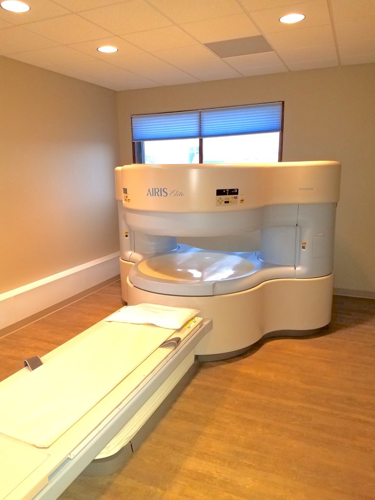
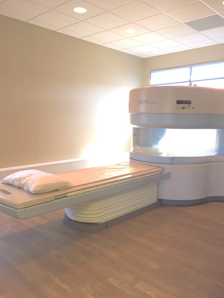
Our HITACHI AIRIS ELITE OPEN MRI is design specifically for larger patients up to 500 pounds. Our machine is also designed to help patient struggling with claustrophobia. This unit is open on the sides to give patients the feeling of openness rather than other MRI units that are tube shaped and patients may have the feeling of being in a pipe or barrel. Also, our room is designed with exterior windows for natural lighting that adds to more of an open feeling helping to combat claustrophobia as well. Most MRI units are in dark rooms with no exterior views. With our HITACHI open unit we are able to put a loved one or family member in the room with the patient to hold her hand during the scan if necessary. The picture below depicts the openness of the room and its natural lighting.